Archive for the ‘Microbe Health Effects’ Category


MICROBIOLOGY FOR THE UNINITIATED – PART 1: WHAT ARE MICROORGANISMS AND WHY SHOULD WE CARE?


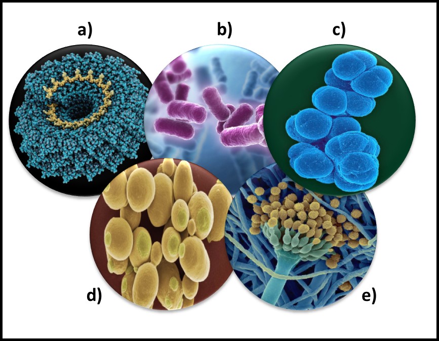
Microorganisms – a) virus; b) bacteria; c) archaea; d) yeast; d) mold.

Each year, on the first morning of STLE’s Metalworking Fluid Management Certificate Training Course, I ask attendees about their backgrounds. Invariably, the majority have at least undergraduate degrees in chemistry, engineering, or chemical engineering. However, of the more than 600 people who have taken the course since 2004, fewer than 50 have had a microbiology course.

Globally, biodeterioration – damage that organisms cause – has been estimated to range between $100 billion and $500 billion annually. Of this, 70 % to 80 % of biodeterioration damage is caused by microorganisms. Add to this an estimated $1 trillion cost due to infectious disease. On the flipside, biotechnology is a $500 billion industry that is based on microbiology. Just considering the economic impact microbe have on our lives, one might reasonably ask why so few people have received the most rudimentary education about microbes. Perhaps it’s the six syllables in microorganism (mic-ro-org-an-is-m) and microbiology that make the topic so intimidating. Still, given the role of microbes in disease, nature’s primary cycles, and biotechnology, one might think that every high school graduate would have learned something about microbiology. In this post and the next several to follow, I will do my best to demystify microbiology.

In this month’s article and those that follow, I’ll offer a very superficial overview of microbiology from an ecological and industrial perspective. There are numerous, excellent introductory microbiology textbooks. I don’t intend to provide any level of detail approaching that of a microbiology textbook. My intention is to help non-technical readers gain a fundamental appreciation for how microbes can affect their lives and their businesses.

Definition

Microorganisms (micro – meaning: small + organism – meaning: something having many related parts that function together as a whole) are living things that are too small to be seen by the naked eye. Microbiologists agree that bacteria, archaea, and some fungi (yeasts and molds) are microorganisms.

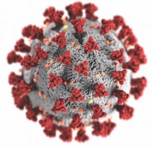
The status of virus is still a matter of debate. Viruses are essentially genetic material packaged in a protein coat. They don’t perform most of the functions that are used to define living beings. Thus most microbiologists do not consider viruses to be life forms. However, viruses reproduce by infecting cells and hijacking their victim’s (host’s) metabolic machinery to reproduce. Consequently, some microbiologists believe viruses should be classified as living things. Image (a) in the title figure shows a tobacco mosaic virus (TMV). The TMV virus is structurally complex, regardless of how we classify it. I included a virus in the in the figure because there are research groups investigating the use of viruses to control microbial contamination in industrial systems. I’ll return to this topic in a future post.

Abundance

One recent study, illustrated in Figure 1, estimated that microorganisms represent ~17% of Earth’s total biomass (estimated total biomass 550 gigatons of carbon – Gt C; 1Gt = 109 tons – and bacterial biomass ≈70 Gt)1. The contribution of bacteria to the Earth’s biomass is second only to that of plants. Microorganism biomass – including that of archaea, bacteria, fungi, protists, and viruses – account for ~93Gt.

A microbiome is the population of all microbes living in a specific ecosystem (fuel tank, cooling tower, human gut, etc.). Researchers investigating the human microbiome have estimated that the average human body has between 1x to 10x as many microbial cells as human cells. Moreover, it has been discovered that tissues, long thought to be microbe-free, have specific microbiomes that are likely to be essential for healthy tissue function (we have known about skin and gut microbes for nearly 170 years, but finding microbiomes specific to nearly every human tissue type (organs, muscle, etc.) was a surprise. Investigators have only scratched the surface of human microbiome research. We have little idea of how microbes interact with human cells and what roles they play in maintaining good health.


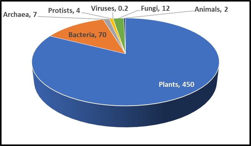
Fig 1. Relative abundance of Earth’s lifeforms, in Gt.

Diversity

As I will explain in future articles, the microbial world is remarkably diverse. The number of different types of bacteria has been estimated to range from hundreds of thousands to tens of millions, of which only a fraction of a percent has been identified.

I’ll discuss why below. Despite their central importance to life as we know it, even the most rudimentary discussion of microorganisms or microbiology (i.e., the study of microorganisms) is rarely included in high school or university curricula.

A Brief History

Tree of Life

Microbes were the first organisms to exist on earth. Our current understanding is that Earth formed 4.6 billion (4,600,000,000) years ago. There is evidence that the first microbes came into existence approximately 3.5 to 3.8 billion years ago (the fossil record indicates that mammals appeared 65 million years ago, and humans showed up a mere 315,000 years ago). Figure 3 illustrates the life on Earth timeline. The bar illustrating Earth’s age is 5 inches (in) long and the one for microorganisms is 4.1 in long. By comparison, the one for plants is 0.2 in and the one for humans is 0.000003 in – too thin to see! Thus, for more than 3 billion years, microbes were the only life forms on Earth.

If we visualize life as a tree, the organisms began to diversify genetically approximately 3.2 to 3.5 billion years ago (Figure 3). This was the time of the last universal common ancestor – LUCA – of all cellular organisms, starting with the bacteria and (thus far) culminating in humans. Before the microbes now classified as members of the kingdom Archaea were discovered near ocean floor, thermal vents in the Marianas Trench, in 1960, the tree of life was thought to have three Kingdoms: Monera (Prokaryota – all single cell organisms that do not have a nucleus or other membrane-bound internal bodies, Protista – all single cell organisms with a nucleus and other membrane-bound bodies, and Eukaryota – all multicellular organisms). As depicted in Figure 3a, in the 1960s through 1990s, Archaea were classified as Archaebacteria. When viewed through a microscope, they appeared to be bacteria. Believing that conditions around Marianas Trench thermal vents was similar to those of primordial Earth, microbiologists initially assumed that Archaea were more ancient than true bacteria – Eubacteriales. As genetic tools became available in the early 1990s, it became apparent that a) Eubacteriales are more ancient than Archaea (Figure 3b), and b) the Archaea are sufficiently distinct genetically to be its own phylogenic Kingdom (phylogenics – the study of evolutionary development and diversification of a species or group of organisms). When Figure 3a was created, fungi were thought to be much more ancient than plants or animals. As Figure 3b illustrates, the phylogenetic tree of life branched off to fungi, plants, and animals a mere half-billion years ago. Thus, fungi are more closely related to us than they are to bacteria.


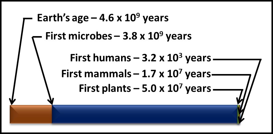
Fig 2. Timelines – microorganisms appeared approximately 1 billion years after Earth was formed and more than 3 billion years before the first plants appeared. Brown bar – Earth’s age; blue bar microorganisms’ age; green bar – time since plants first appeared; yellow bar – time since first animals appeared; purple bar (invisibly narrow line) – time since humans appeared.


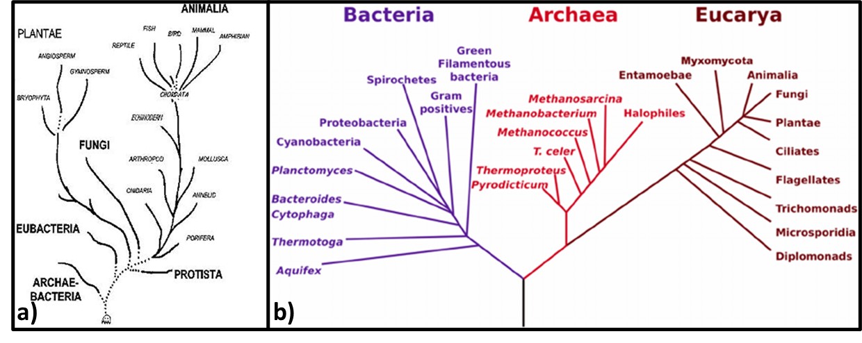
Fig 3. Phylogenic trees – a) Tree from mid-1960s depicting Archaebacteria as being more ancient than Eubacteria; b) Woese et al. (19902) phylogenic tree showing three Kingdoms – Bacteria, Archaea, and Eucarya. Line segment lengths are based on genetic differences (longer lines indicate greater differences). The initial split at bottom center is LUCA.

Genomic testing has been a cottage industry since the late 1990s. A recent diagram (Figure 4) is based on the genomics of 2.3 million different species, from bacteria to humans, illustrates how complex the Tree has become (or as some authors note, the Tree now looks more like a Bush).


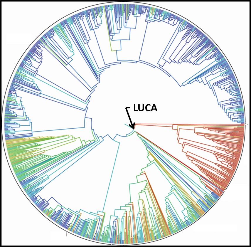
Fig 4. 2015 Tree of Life based on genetic data from 2.3 species.3

Human Awareness

Humans have been using microbes from time immemorial. We have been fermenting grains and grapes, and making cheeses, for as long as we have been cultivating plants or maintaining livestock. Modern microbiology dates from 1665, when Roert Hooke published Micrographia: or some Physiological Descriptions of Minute Bodies made by Magnifying Glasses with Observations and Inquiries Thereupon. A decade later, Antonie van Leeuwenhoek published his observations. Hooke and van Leeuwenhoek had each constructed microscopes through which they observed and created sketches of microorganisms (Figures 5a and 5b). However, it took another 200 years before Louis Pasteur and Robert Koch demonstrated that microbes were living beings and were responsible for fermentation, disease, and spoilage.


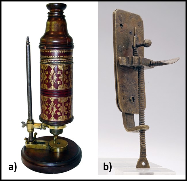
Fig 5. First microscopes – a) replica of Robert Hook’s microscope; b) replica of Antonie van Leeuwenhoek’s microscope.

Louis Pasteur conducted experiments to disprove the theory of spontaneous generation (belief that microbes developed from inanimate components of the materials which they caused to rot) and prove the germ theory of disease (i.e., that microorganisms did not form spontaneously and that they caused disease, biodeterioration, and were the agents responsible for fermentation). Pasture used goosenecked flasks (Figure 6) to demonstrate that microbes did not proliferate (multiply) in broth boiled in the flasks but did in identical flasks that contained unboiled broth. Proliferation also occurred when Pasteur intentionally permitted boiled broth to be exposed to microbially contaminated air (i.e., either by breaking off the gooseneck or tipping the flask). The gooseneck shape allowed air but not microbes to enter the flasks. These experiments led to Pasteur’s development of the pasteurization – the process of heating substances at temperatures sufficient to disinfect but not degrade them.


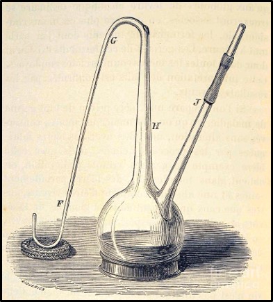
Fig 6. Drawing of Louis Pasteur’s gooseneck flask used to disprove spontaneous generation theory.

While Pasteur was focusing on fermentation microbiology, Robert Koch demonstrated the unequivocal relationship between microbes and disease. Koch demonstrated that the disease, anthrax was caused by the spore forming bacterium, Bacillus anthracis. He also developed the first solid growth media so that he could isolate pure cultures from colonies (Figure 7, zone 5). Pasteur’s and Koch’s research launched the modern age of microbiological research.


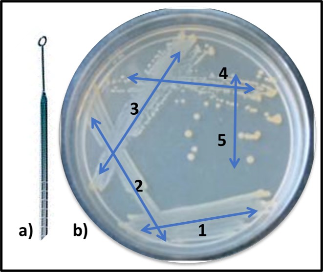
Fig 7. Obtaining a pure (single type of bacterium) culture by the streak plate method – a) specimen is collected using a sterilized inoculating loop; b) successively, initial specimen is deposited onto solid nutrient medium using a back-and forth motion (1), inoculating loop is heat sterilized, cooled, and oved across initial inoculation zone (2). This dilutes the sample. The final iteration (5) typically produces individual colonies after the inoculated plate has been incubated.

As now, much of the research performed during the late 19th century was focused on the relationship between microbes and disease. However, Sergei Winogradsky became the father of microbial ecology. Based on his pioneering research on sulfur metabolism in the late 1880s, Winogradsky developed the theory of biogeochemical cycles. This theory states that elements like sulfur, carbon, nitrogen, and phosphorous cycle through nature (Figure 8). These cycles are primarily mediated by microbial activity. The first paper describing gasoline deterioration by microbes was published in 1895. Starting in the 1920s, considerable effort was focused on oilfield damage caused by microorganisms. This research included the first studies on what was originally called microbially induced corrosion (MIC – now microbiologically influenced corrosion – see MICROBIOLOGICALLY INFLUENCED CORROSION – Biodeterioration Control Associations, Inc. (biodeterioration-control.com)). However, the term biodeterioration was not coined until 1965, when H. J. Hueck offered the definition: “any undesirable change in the properties of a material caused by the vital activities of organisms.”4


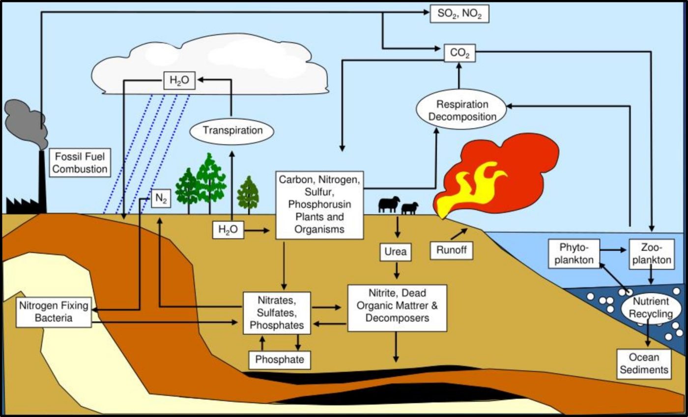
Fig 8. Biogeochemical cycles – this illustration provided a simplified depiction of how carbon (C), nitrogen (N), phosphorous (P), and sulfur (S) cycle through nature.

In my seminars on the topic I explain that biodeterioration and bioremediation are two sides of the same biodegradation coin (Figure 9). As Winogradsky observed, microbes drive biogeochemical cycles. These cycles occur regardless of human intent. When we want biodegradation to occur, we call it bioremediation. When microbial activity causes changes, we’d prefer to prevent, we call it biodeterioration (see https://biodeterioration-control.com/2018/03/).


Fig 9. Like the obverse (front) and reverse (back) sides of this 2000 Sacagawea U.S. dollar coin, bioremediation and biodeterioration are flips sides of biodegradation.

Summary

Microbes play invaluable roles in our lives. Our bodies would not function without the microbes that make up he human microbiome. Fewer than 2,000 pathogenic microbes have been identified among the tens of thousands that have been identified and the millions of different types of microbes that exist in nature. Microbes mediate nutrient cycles. This cycling conserves essential nutrients and prevents wastes from accumulating. Biodegradation includes all processes that breakdown organic substances. On one hand, biodegradation is the foundational element of a $0.5 trillion biotechnology industry. On the other, biodeterioration and infectious disease cost $1.5 trillion per year. We are part of the microbial world. To me, that seems like an excellent reason why everyone should have a basic understanding of microbiology.

For more information about fuel system condition monitoring and predictive maintenance, contact me at fredp@biodeterioration-control.com.


1 Bar-On, Y.M, Phillips, R., Milo, R., 2018. The biomass distribution on Earth. Proc Natl Acad Sci U S A., 115(25):6506-6511. https://pubmed.ncbi.nlm.nih.gov/29784790/.
2 Woese, C. R., Kandler, O., Wheelis M.L., 1990. Towards a natural system of organisms: proposal for the domains Archaea, Bacteria, and Eucarya. Proceedings of the National Academy of Sciences of the United States of America. 87 (12): 4576–9. https://doi.org/10.1073/pnas.87.12.4576.
3 Hinchliff, C. et al., 2015. Synthesis of Phylogeny and Taxonomy Into a Comprehensive Tree of Life. Proceedings of the National Academy of Sciences https://www.pnas.org/doi/full/10.1073/pnas.1423041112.
4 Hueck, H.J., 1965. The Biodeterioration of Materials as a Part of Hylobiology. Material und Organismen, 1, 5-34.

Minimizing Covid-19 Infection Risk In The Industrial Workplace


Electron microscopy image of the SARS-CoV-2 virus.


COVID-19 Infection Statistics

Although anti-COVD vaccines are rolling out and people are being immunized, as of early late December 2020, the rate at which daily, newly reported COVID-19 cases has continued to rise (Figure 1). In my 29 June 2020 What’s New article I discuss some of the limitations of such global statistics. In that post, I argued that the statistics would be more meaningful if the U.S. Centers for Disease Control’s (CDC’s) morbidity and mortality reporting standards were used. Apropos of COVID-19, morbidity refers to patients’ cases reported and having the disease and mortality refers to COVID-19 patients who die from their COVID-19 infection. Both morbidity and mortality are reported as ratios of incidence per 100,000 potentially exposed individuals. I illustrated this in my portion of an STLE webinar presented in July 2020.


Fig 1. Global incidence of new COVID-19 cases – daily statistics as of 23 December 2020 (source: coronavirusstatistics.org).


What Do the Infection Statistics Mean?

Social scientists, epidemiologists, and public health specialists continue to debate the details, but the general consensus is that the disease spreads most widely and rapidly when individuals ignore the fundamental risk-reduction guidelines. It appears that COVID 19 communicability is proportional to the number of SARS-CoV-2 virus particles to with individuals are exposed. Figure 2 illustrates the relative number of virus particles shed during the course of the disease.


Fig 2. Relationship between number of SARS-2CoV viruses shed and COVID-19 disease progression.


Notice that the number of viruses shed (or dispersed by sneezing, coughing, talking, and breathing) is quite large early on – before symptoms develop fully. It’s a bit more complicated than that, however. Not all infected individuals are equally likely to shed and spread the virus. All things being apparently equal, some – referred to as super-spreaders – are substantially more likely than others to infect others. Although people with or without symptoms can be super-spreaders, those who are infected but asymptomatic are particularly dangerous. These folks do not realize that they should be self-quarantining. A study published in the 06 November 2020 issue of Science (https://science.sciencemag.org/content/370/6517/691) reported that epidemiological examination of millions of COVID-19 cases in India revealed that 5 % of infected people were responsible for 80 % of the reported cases.

What Shall We Do While Waiting for Herd Immunity to Kick-In?

The best strategy for avoiding the disease is to keep yourself physically distanced form others. Unfortunately, this advise is all but worthless for most people. We use public transportation to commute to work. We teach in classrooms, work in offices, restaurants, medical facilities, and industrial facilities in which ventilation systems are unable to exchange air frequently enough to minimize virus exposure risk. The April 2020 ASHRE Position Document on Infectious Aerosols recommends the use of 100 % outdoor air instead of indoor air recirculation. The same document recommends the used of high-MERV (MERV – minimum efficiency removal value – 10-point scale indicating the percentage of 0.3 µm to 10 µm particles removed) or HEPA (HEPA – high efficiency particulate absorbing – able to remove >99.9% of 0.3µm particles from the air) filters on building HVAC systems. Again, as individuals who must go to work, shop for groceries, etc., outside our own homes, we have little control over building ventilation systems.

Repeatedly, CDC (Centers for Disease Control), HSE (UK’s Health and Safety Executive), and other similar agencies have offered basic guidance:

1. Wear face masks – the primary reasons for doing this is to keep you from transmitting aerosols and to remind you to keep your hands away from your face. Recent evidence suggests that that although masks (except for ones that meet N-95 criteria) are not very efficient at filtering viruses out of the air inhaled through them, they do provide some protection.

2. Practice social distancing to the extent possible. The generally accepted rule of thumb is maintaining at least 6 ft (1.8 m) distance between people. This is useful if you are in a well-ventilated space for relatively short periods of time but might be insufficient if you are spending hours in inadequately ventilated public, industrial, or institutional spaces.

3. Wash hands thoroughly (at least 30 sec in warm, soapy water) and frequently. The objective here is to reduce the chances of first touching a virus laden surface and then transferring viruses into your eyes, nose, or mouth.

Here are links to the most current guidance documents:

CDC – How to Protect Yourself and Othershttps://www.cdc.gov/coronavirus/2019-ncov/prevent-getting-sick/prevention.html

CDC – Interim Guidance for Businesses and Employers Responding to Coronavirus Disease 2019 (COVID-19), May 2020https://www.cdc.gov/coronavirus/2019-ncov/community/guidance-business-response.html

HSE – Making your workplace COVID-secure during the coronavirus pandemichttps://www.hse.gov.uk/coronavirus/working-safely/index.htm

UKLA- HSE Good Practice Guide – http://www.ukla.org.uk/wp-content/uploads/HSE-Good-Practice-Guide-Sept20-Web-LowresC.pdf – discusses health & safety in the metalworking environment.

WHO – Coronavirus disease (COVID-19) advice for the publichttps://www.who.int/emergencies/diseases/novel-coronavirus-2019/advice-for-public

Remember: Prevention really Means Risk Reduction

It is impossible to reduce the risk of contracting COVD-19 to zero. However, timely and prudent preventative measures can reduce the risk substantially so that people can work, shop, and interact with one another safely. Guidance details continue to evolve as researchers learn more about SAR-CoV-2 and its spread. However, the personal hygiene basics have not changed since the pandemic started a year ago. If each of us does our part, we will be able to reduce the daily rate of new cases dramatically, long before the majority of folks have been immunized.

For more information, contact me at fredp@biodeterioration-control.com

REMEMBERING A MENTOR AND A MENSCH – PROFESSOR EUGENE D. WEINBERG 1922 TO 2019

This morning, while reading the Fall 2019 issue of Indiana University Alumni Magazine, I was saddened to read Gene Weinberg’s name in the list of recently deceased IU faculty and staff.
Professor Emeritus Eugene D. Weinberg died on 08 March – less than a week after having celebrated his 97th birthday. Gene was the first academician to have had a profound effect on my life’s path. I know that his memory will be a blessing to all of us who had the privileged and pleasure of knowing him.

I first met Professor Weinberg in 1966 – a few weeks into my first semester at IU. My initial plan was to have been a math major, but within a month, I began to rethink that plan. Having been tinkering with microbiology since my parents made the mistake of presenting me with a microscope for my eighth birthday, I decided to explore the possibility of changing majors to microbiology. In late October 1966, I visited with Professor Weinberg in his Jordan Hall office to explore my options. He advised me that the courses that I was taking were perfectly aligned with those that would be part of a microbiology major. He contacted my original, math department advisor and agreed to become my faculty advisor. From that date through my graduation in June 1970, Gene was always available to offer guidance and to facilitate my efforts to perform extracurricular studies under various Microbiology Department professors. Although I never saw it, I have no doubt that Gene’s letter of recommendation helped me to get accepted into graduate school and receive a full fellowship for my studies at University of New Hampshire.

Gene’s research interest was in medical microbiology. Knowing that my passion was microbial ecology, while I was taking his course in Medial Microbiology, he encouraged me to make my class project ecologically focused. When I went home for Thanksgiving, 1968, I took a suitcase full of sterile, 100 mL glass bottles with me. One the Friday after Thanksgiving, I drove to the Delaware River’s source. From there, and at various bridges located at 50 mi intervals – ending at the Delaware Memorial Bridge, I used a fishing pool, jury-rigged sampling setup to collect samples from each bank and the middle of the river. I then carried the full bottles back to Bloomington (good thing this was before there were suitcase weight limitations or TSA) where I proceed to run culture tests and biochemical taxonomic profiles on each type of microbe that I had detected. I rationalized this survey effort by noting that there was a possibility that the taxonomic profiles along the river’s length might have been indicative of public health risks.

I didn’t realize it at the time, but that project marked the start of my career as a microbial ecologist. I did realize from the outset that Gene was a supportive, encouraging mentor. When others might have said: “you can’t do that!” Gene would always tell me that I had a great idea, asked me if I had thought about various details – which of course I hadn’t, and suggest research papers that might help me to refine my thoughts. Gene was one of perhaps four mentors whose influence shaped my career as a microbiologist. I feel most fortunate for having known him and have benefited from his wisdom, his kindness, and his mentorship.

You can find Gene’s full obituary article at https://www.hoosiertimes.com/herald_times_online/obituaries/eugene-weinberg-phd/article_f86ed715-7dde-5789-9916-40d1e0fb0bfe.html.

METAWORKING FLUIDS, 3RD EDITION NOW AVIALABLE!

Thirteen years after Metalworking Fluids, 2nd Ed. was published, the third edition is now available. Metalworking Fluids, 3rd Ed. Jerry Byers, Ed. has just been published (ISBN, Hardbound: 978-1-4987-2222-3; E-book: 978-1-14987-2223-0) and is available from STLE, CRC Press, or Taylor & Francis.

MWF 3rd. Ed. promises to become the new MWF bible. All of its chapters reflect either substantial updates or all new material. I recommend this new volume most strongly to all metalworking industry stakeholders.

Full disclosure, I wrote Chapter 11 – Microbiology of Metalworking Fluids. Many of the other chapters were written by colleagues on STLE’s Metalworking Fluid Education and Training Subcommittee.

The Truth is Out There…

For those of you who are interested in metalworking fluid microbiology and microbial contamination control, I invite you to read my March 2016 Tribology and Lubrication Transactions TLT) article: MWF Biocides Part II – Science vs. Fiction.
This was an accidental article that I was asked to write in response to an error-laden article that had appeared in TLT’s November issue. The earlier piece had been written by an individual whose familiarity with the topic was limited to the research performed in the process of drafting the TLT submission. I had not yet read the article when I started receiving flaming emails from industry colleagues who mistakenly believed that I had an editorial role and had somehow approved the article for publication. Initially, my plan was to write a letter to the editor. Indeed, I wrote a draft letter listing each error and the correct information (with relevant references cited as appropriate). The letter morphed into the March article. To be sure that I wasn’t just offering my personal opinions, I recruited log time colleagues Drs. Neil Canter and Alan Eachus and Mssrs. Jerry Byers and Richard Rotherham to co-author the article. I am much indebted to each of them for their contributions to the effort.
MWF Biocides Part II focuses primarily on the scientifically unsupportable conflation of formaldehyde (HCHO) and formaldehyde-condensate microbicides (FCM). The toxicological profiles of FCM differ among specific chemistries, but as a group are substantially different from HCHO. Moreover, although regulators assume that 100% of the HCHO in FCM will end up in the air above metalworking fluids (MWF) threated with FCM, data prove otherwise. Over the past couple of years, the number of microbicides approved for use in MWF has plummeted. In Europe there are only 27 listed biocidal substances (most are still going through regulatory review) that can be used in MWF. In the U.S., by last summer, the US EPA’s Office of Pesticides Programs will most likely issue guidance that will determine the future availability of FCM. In addition to clarifying the FCM issues that had been misreported in the November article, the March article sets the record straight on nearly 30 other misstatements made in the earlier publication.
Please contact me at fredp@biodeterioraiton-control.com for a copy of the MWF Biocides Part II.

Legionella pneumophila in Metalworking Fluids

I’m sharing an email exchange that I had with a colleague who had asked about the risk of L. pneumophila (the microbe that causes Legionnaire’s disease) in MWF.
Thank you for posting your query to BCA’s website.

You wrote:
“I wondered if you could help me answer a customer’s question. One of my customer’s machine tool operators is in the hospital being treated for Legionnaires’ disease. My customer asked me if the Kathon 886 MW or Kathon CC kills this strain of bacteria. I really appreciate your help and advice. I attend the annual STLE meeting every year and hear you speak on maintaining and monitoring metal working fluids, so I thought you would be the best source to ask. The Legionnaires’ disease was most likely contracted in Tennessee while this gentleman was on vacation. Other machine operators are now afraid they might contract the disease through the metal working fluids in the plant.
Thank you for your time and thoughts.”

The short answer is yes.

Not long after Legionella pneumophila was identified as the disease agent that caused Legionnaire’s disease, Rohm & Haas tested Kathon WT1.5 efficacy against the bacterium. WT1.5 is just Dow’s (formerly R & H) water treatment market label for the 1.5% active product we use as Kathon 886MW and 886MW 1.5 in the MW industry.

Keep in mind that L. pneumophila is ubiquitous. If you recall the incident at Ford’s Le Brea, OH plant some years ago, four machinists came down with Legionnaire’s disease. Attempts to detect L. pneumophila from MWF systems all failed. An immunological survey of all of the plant’s employees revealed that the majority has antibodies to L. pneumophila. Other immunological surveys (populations outside our industry) have demonstrated that the majority of the population has been exposed to the microbe (i.e.: has the antibodies). Most of the time, folks who contract the disease have other health problems that render them more susceptible than the general population. Back to Le Brea. That incident and a cluster of Pontiac Fever cases at a Pontiac Plant in Windsor Ontario in 1981 are the only two clusters of Legionnaire’s disease that have been reported in the MW industry. The 1981 outbreak was caused by L. feeleii growing in the facility’s cooling towers. The source of L. pneumophila at Le Brea was never confirmed.

From what we know, workers are much more likely to be at risk from improperly controlled heat exchange systems/cooling towers than from MWF.

OUR SERVICES

  • Consulting Services
  • Condition Monitoring
  • Microbial Audits
  • Training and Education
  • Biocide Market Opportunity Analysis
  • Antimicrobial Pesticide Selection

REQUEST INFORMATION


    captcha